Fat breasts how do they affect breast cancer?
Periskopi (4 years ago)
The bolt on what the dense breast tissue means, how a doctor estimates which woman has thick breasts, how breast density affects mammography, and what additional tests are available to detect dense breast cancer in the breast tissue refers to [...]
The bolt on what the dense tissue of the breast means, how a doctor estimates that a woman has thick breasts, how breast density affects mammography, and what additional tests are available to detect breast cancer
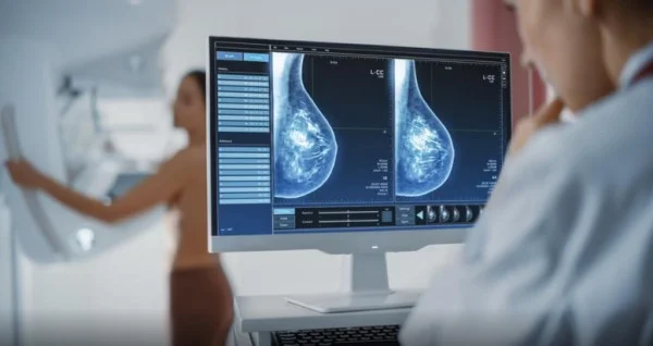
The dense breast tissue refers to the appearance of the breast tissue in the mammography, which is a normal and common find. The breast tissue is made up of milk glands, milk canals, supportive tissue, and fat tissue. When looking at a mammography, thick - breasted women have a thicker tissue than fat that looks dark and transparent in mammography.
The dense breast tissue appears as a dense white surface in the mammography, making it difficult to see any change. The tissue density depends on the number of cells, and more cells mean higher density. The breast gland India, which produces milk, contains a higher concentration of cells, so younger women have higher breasts.
Because dense breast tissue makes reading mammographics difficult, since it is harder to detect all the bumps and areas of abnormal tissue, younger women are recommended to an ultrasound of the breast instead of mammography. With aging, the tissue in the glands shrinks, replacing it with the fat tissue and thus becoming more transparent for mammography.
How do doctors determine if you have thick breasts?
Radiologist who analyzes mammography determines the ratio of fat and dense tissue and determines the degree of breast density. The Gulf Image Reporting and Data System (BI-RADS) is used to describe density levels. The British Mayo Clinic explains that levels of density in the mammography density report fall to four categories marked by ACR A, B, C and D:
ACR Aʹ shows that the chest is almost entirely composed of fat tissue (up to 25% density). On average, one in 10 women has such breasts.
ACR B shows that there are dense areas (up to 50% density), but most of the bay is not dense. On average, 4 out of 10 women have such a finding.
ACR C shows there are areas of density of between 51 and 75%. On average, 4 out of 10 women have such a finding.
ACR D (extraordinaryly dense) means that almost all breast tissue is dense (> 75%). On average, one in 10 women has such breasts. In such structured breasts, mammography must be complete with ultrasound.
Fat breasts are thought to have women whose breasts are regarded as thick or extremely thick ( ACR C and D). About 50% of women making mammography have thick breasts.
What can cause dense breast tissue?
It is not clear why some women have a lot of dense tissue, and others cannot be affected. You're more likely to have fat tits if:
You're the youngest. The breast tissue tends to get less dense with age, although some women may have dense breast tissue at any age.
You have a lower body mass index. Low - fat women are more likely to have denser breast tissue than women who are obese.
You're getting hormone therapy in menopause. Women who receive combined hormonal therapies to alleviate symptoms of menopaus are more likely to have thick breasts.
Why is chest density important?
Fat breasts may influence:
They increase the likelihood that a mammography will not detect breast cancer, as dense breast tissue may mask possible differences.
They increase the risk of breast cancer, although doctors are not sure why.
What tests are recommended to detect breast cancer?
Most medical organisations recommend that women, with an average risk of breast cancer, begin regular mammograms starting at the age of 40 and repeat them each year. Women with thick breasts but without other risk factors for breast cancer are thought to have a higher risk than the average breast cancer. The dense breast tissue makes it difficult to interpret mammography, since breast cancer and dense tissue appear in mammography as a white surface. Very thick breasts can increase the risk of cancer not being discovered in mammography.
Despite concerns about the discovery of cancer in dense breasts, mammography remains an effective test. The most common type of mammography, digital mammography, preserves bay images as digital files instead of film files and allows more detailed analysis. This is a more effective method of finding cancer in dense breast tissue than the oldest mammography technology in film.
Are other research more effective?
There is evidence that additional tests can increase the likelihood of the discovery of cancer in dense breast tissue. However, they carry additional risks and have not proved that an additional method of testing reduces the risk of breast cancer mortality. You can discuss with your doctor the need for additional tests based on other risk factors.
Additional tests to detect breast cancer may include:
3-D mammography. Tomosynthesis uses ionistic X-rays to collect multiple bay images from different directions, and images connect the computer to a 3D view of the bay.
FRIENDS. The breast's magnetic resonance does not use ionizing radiation. High - risk women are advised to develop breast cancer, such as women with genetic mutations that increase the risk of developing breast cancer or women who have been shot down in the chest wall area.
Bay ultrasound. Ultralingul uses mechanical waves to analyze tissues. Diagnostic ultrasound is often used to evaluate suspicious areas discovered in mammography. Aside from mammography, an ultrasound examination of increased genders and density is recommended to be filled with ultrasound, which is also a common breast examination of pregnant women and women up to 40 years old.
The quality of the examination depends on the experience of the person performing it
Medieval breast images (MBI). The breast's molecular image uses a special gamma camera that records the activity of radio reliefs applied to a vein in the arm. Normal tissue and cancer tissue collect different radiods, which can be seen in images on gamma cameras.
Each approach has advantages and disadvantages, but although each test has shown that it detects more breast cancer than a mammography, none of these newest imaging tests have been shown to reduce the risk of breast cancer mortality, unlike a standard film mamography.